
TL;DR

Turn quiet afternoons into listening time
- Thousands of audiobooks, podcasts and originals
- Listen on your phone, tablet or Echo — also offline
- Cancel anytime
A paper in Nature Communications describes the Knight Alzheimer Research Imaging dataset, which includes 6,217 brain-imaging acquisitions from 1,645 people aged 42 to 97. The resource combines MRI, amyloid and tau PET scans with cognitive and clinical information, offering researchers a way to investigate associations across healthy aging and Alzheimer’s disease—not proof that the scans predict or prevent the disease.
A paper published in Nature Communications describes a research dataset containing 6,217 brain-imaging acquisitions from 1,645 people aged 42 to 97, giving scientists a resource to examine how brain structure and Alzheimer’s-associated proteins relate to cognition over time. The Knight Alzheimer Research Imaging dataset combines MRI and amyloid and tau PET scans with clinical and cognitive assessments; it is a research resource, not a test that diagnoses or predicts Alzheimer’s disease.
The dataset, known as KARI, was developed by the Knight Alzheimer Disease Research Center at Washington University in St. Louis. It includes people across a range from healthy aging and preclinical Alzheimer’s disease to symptomatic disease. More than 800 participants had imaging at multiple time points, and some were followed for more than 15 years. The paper describes the resource and examines associations between imaging measures, age, clinical impairment and cognitive performance.
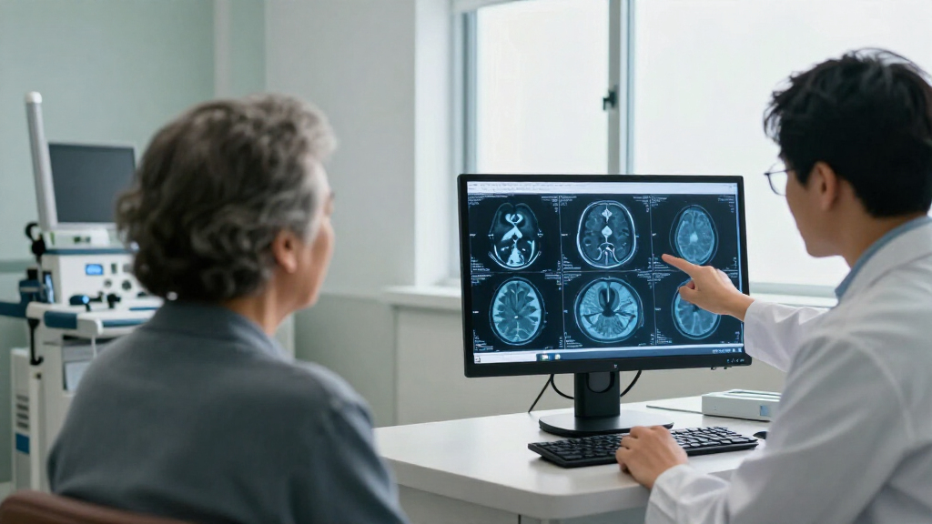
The imaging covers different aspects of the brain. Structural, functional and diffusion MRI provide measures of brain anatomy and tissue, while PET scans assess amyloid and tau, proteins associated with Alzheimer’s. The collection includes 3,385 MRI sessions, 2,684 amyloid PET scans and 1,113 tau PET scans. A total of 792 participants had all three imaging types, allowing researchers to compare measures in the same people.
Clinical and cognitive information adds another layer. Assessments were available for 1,643 participants within two years of their scans. Most participants had a maximum Clinical Dementia Rating score of 0, indicating no impairment on that scale; 362 reached 0.5, 150 reached 1 and 11 reached 2. The dataset also includes genetic information, fluid biomarkers and demographic details. Qualified researchers can access raw imaging and processed measures, including quantified biomarkers and anatomical segmentations.
A Longer View of Early Brain Changes
Alzheimer’s-related changes can begin years or decades before noticeable symptoms, according to the research paper. Repeated scans paired with cognitive assessments may let researchers study how amyloid, tau and brain measures vary alongside cognitive health, including among people who do not yet show clinical impairment. That timing matters because research into the preclinical period may help clarify when and how changes emerge.
The value is in the combined, longitudinal record—not simply the number of scans. Researchers can examine several imaging types and clinical measures together, and revisit some participants over time. The study reports associations between imaging biomarkers and clinical or cognitive status; it does not establish that any one scan causes, predicts or confirms an individual’s future decline. Nor does describing the dataset demonstrate that a treatment can prevent or cure Alzheimer’s.
Alzheimer's disease brain health books for general readers
As an affiliate, we earn on qualifying purchases.
As an affiliate, we earn on qualifying purchases.
How the KARI Resource Was Built
The KARI resource comes from a single research center, where uniform collection standards can support consistency across participants and visits. The paper notes that some variation in imaging hardware is inevitable, a consideration for scientists analyzing measurements collected over an extended period.
The cohort is not evenly divided by clinical status. Most participants had no impairment recorded, and the groups differed in age and other characteristics: participants with a Clinical Dementia Rating above 0 averaged 75.7 years, compared with 70 years among those scoring 0. The paper also reports differences in education, sex distribution and APOE ε4 carrier frequency between the groups. These factors are relevant when interpreting associations, and the authors’ cognitive analyses adjusted for age, education, sex and APOE ε4 status.
“The dataset is intended to provide researchers with “the necessary context and quality-assurance metrics to utilize this expansive resource for their own investigations.””
— The study authors, as described in the paper
brain anatomy and neuroscience atlas book
As an affiliate, we earn on qualifying purchases.
As an affiliate, we earn on qualifying purchases.
Limits of the Scan Findings
The paper describes the cohort and reports relationships between imaging biomarkers and clinical or cognitive measures, but the supplied report does not provide detailed effect sizes or results for each biomarker. The strength and practical meaning of particular associations therefore cannot be judged from the dataset description alone. An association does not show that an imaging change causes cognitive impairment or can forecast what will happen to a specific participant.
The cohort also reflects people enrolled and followed at one research center, and the clinical groups differ in age and other characteristics. While repeated observations and statistical adjustments help researchers examine these data, they do not remove every limitation or establish that findings will apply equally to all populations. The paper does not report a new Alzheimer’s treatment, prevention method or clinical screening recommendation.
memory and cognitive health activity books for adults
As an affiliate, we earn on qualifying purchases.
As an affiliate, we earn on qualifying purchases.
Further Studies Using the Dataset
The immediate next step is for qualified researchers to use the KARI data to investigate questions about brain aging and Alzheimer’s-associated changes. Its combination of repeated imaging, clinical assessments and cognitive measures can support analyses that follow participants across time and compare different imaging biomarkers. The dataset paper provides information intended to help researchers assess data quality and choose measures for their investigations.
Any future claims about early detection, prognosis or treatment will depend on the results of those separate studies and how well they are supported. The report does not specify a next trial, publication date or clinical application. For now, the confirmed development is the publication and description of a research resource—not a change to Alzheimer’s diagnosis or care.
MRI and PET imaging textbook neuroscience
As an affiliate, we earn on qualifying purchases.
As an affiliate, we earn on qualifying purchases.
Key Questions
What is the KARI dataset?
It is a research collection developed by Washington University in St. Louis’s Knight Alzheimer Disease Research Center. It includes imaging and clinical, cognitive, genetic and other information from 1,645 participants.
What kinds of brain scans does it include?
The resource includes structural, functional and diffusion MRI, as well as PET imaging for amyloid and tau. Some participants had multiple imaging types and repeat scans.
Do the scans show who will develop Alzheimer’s?
No such predictive test is established by this paper. It describes a dataset and examines associations between imaging measures and clinical or cognitive status; those associations do not diagnose or predict an individual’s future disease.
Why are repeated scans useful?
Repeated observations can help researchers examine how imaging measures and cognition change within people over time. More than 800 participants had longitudinal imaging, with some followed for more than 15 years.
Can the public access the scans?
The paper says raw imaging and processed measures are available to qualified researchers. It does not describe unrestricted public access.
Source: rss
Cold & flu season Picks
humidifiers
As an affiliate, we earn on qualifying purchases.